-40%
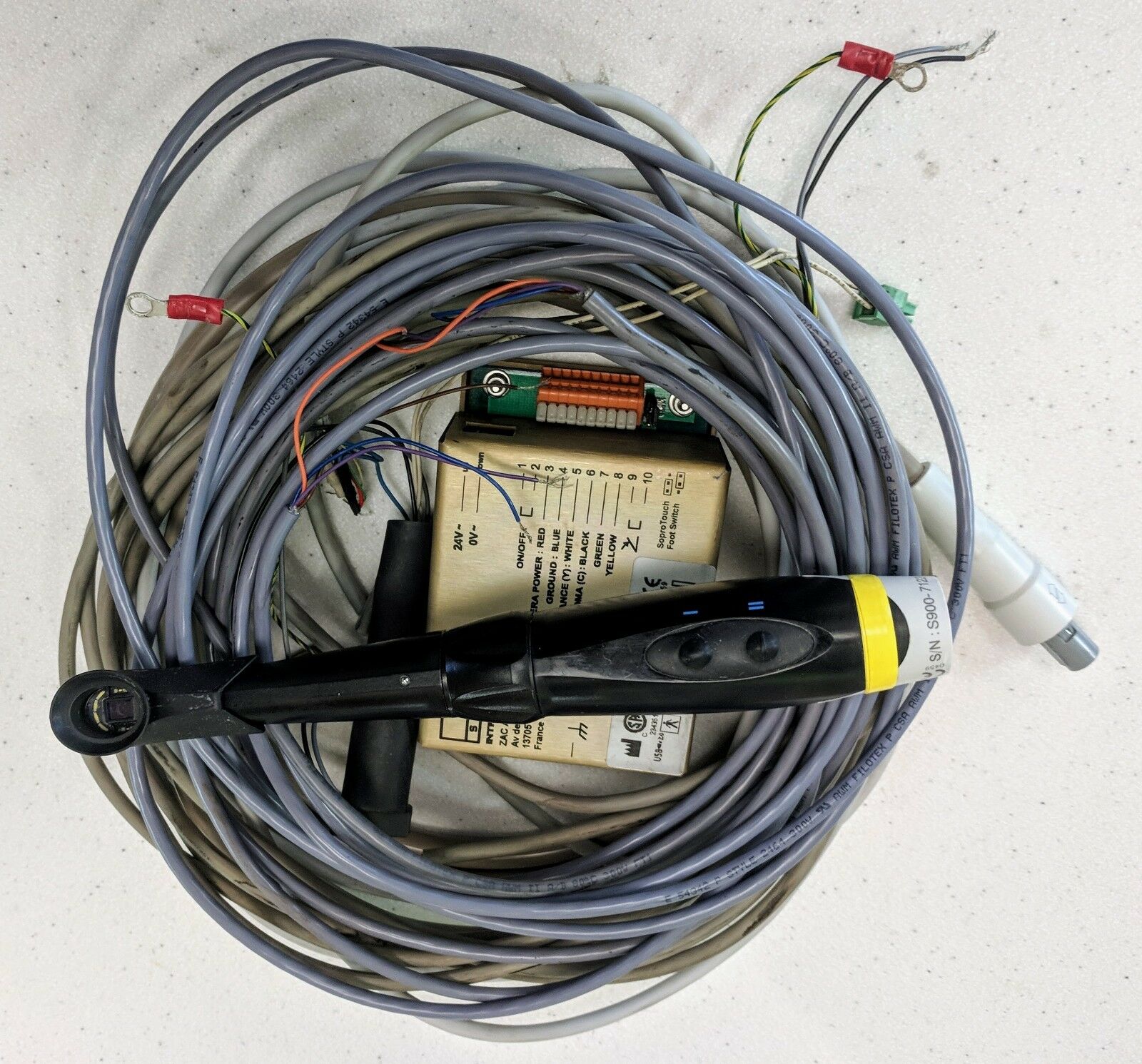
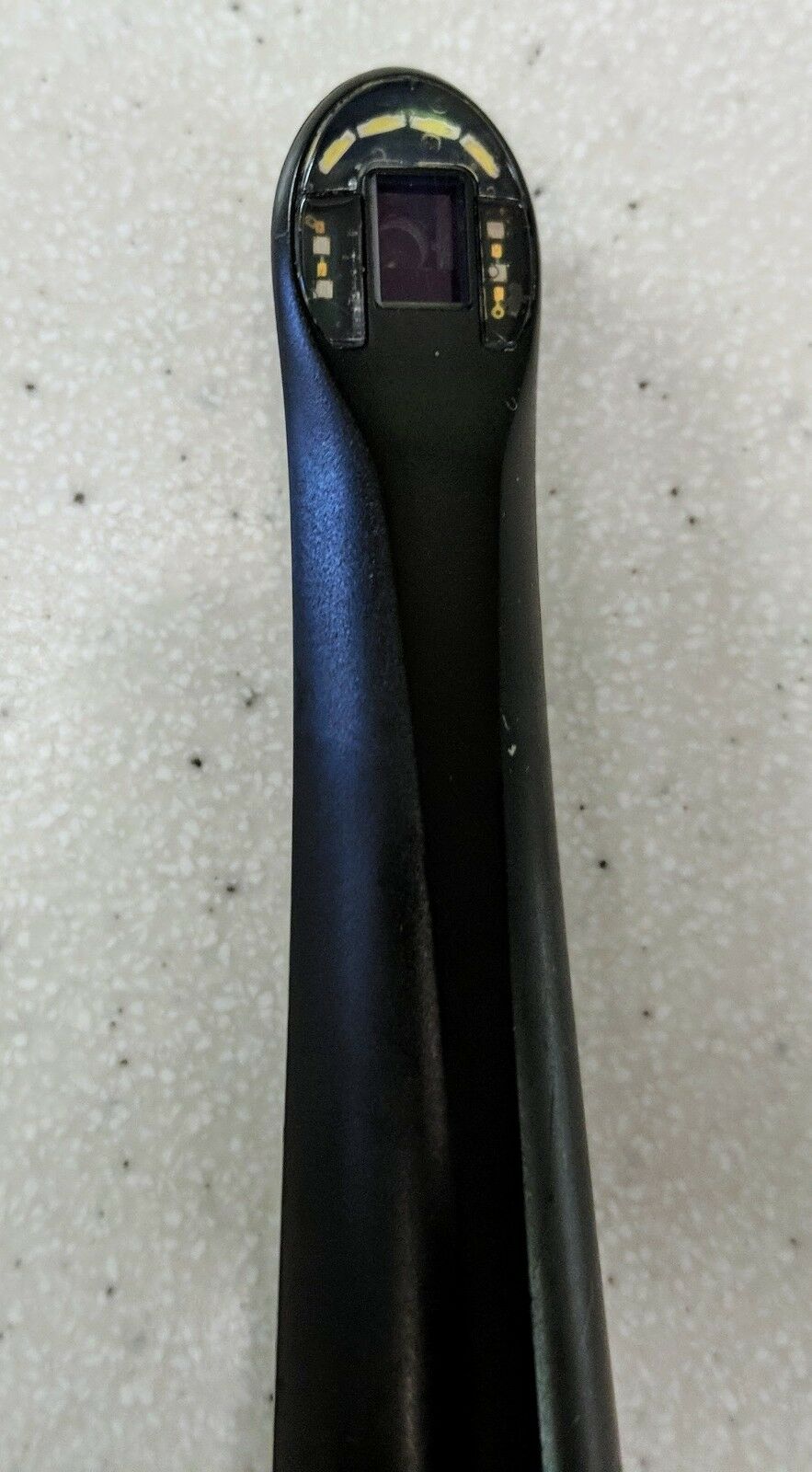
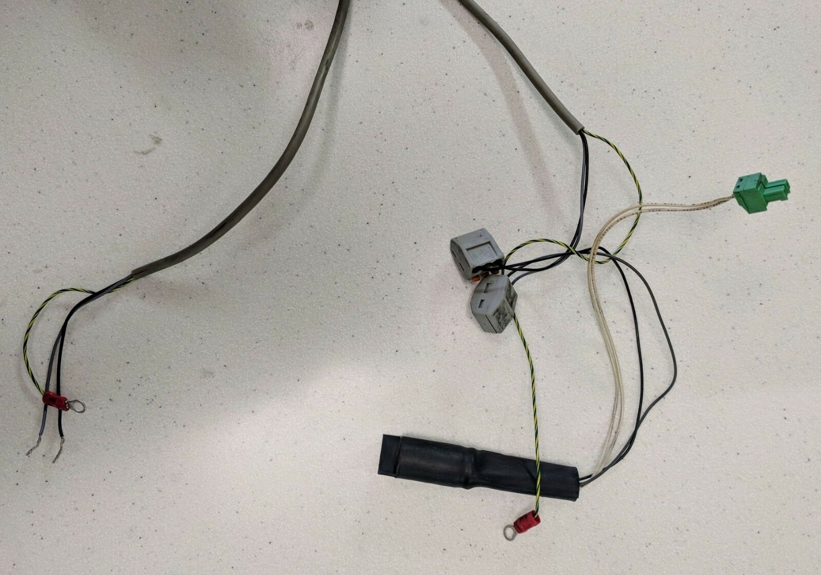
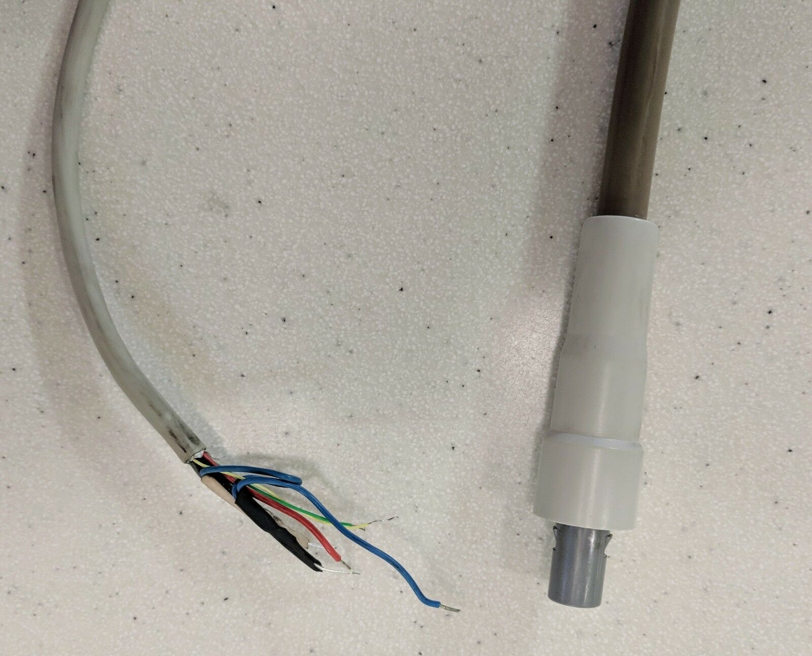
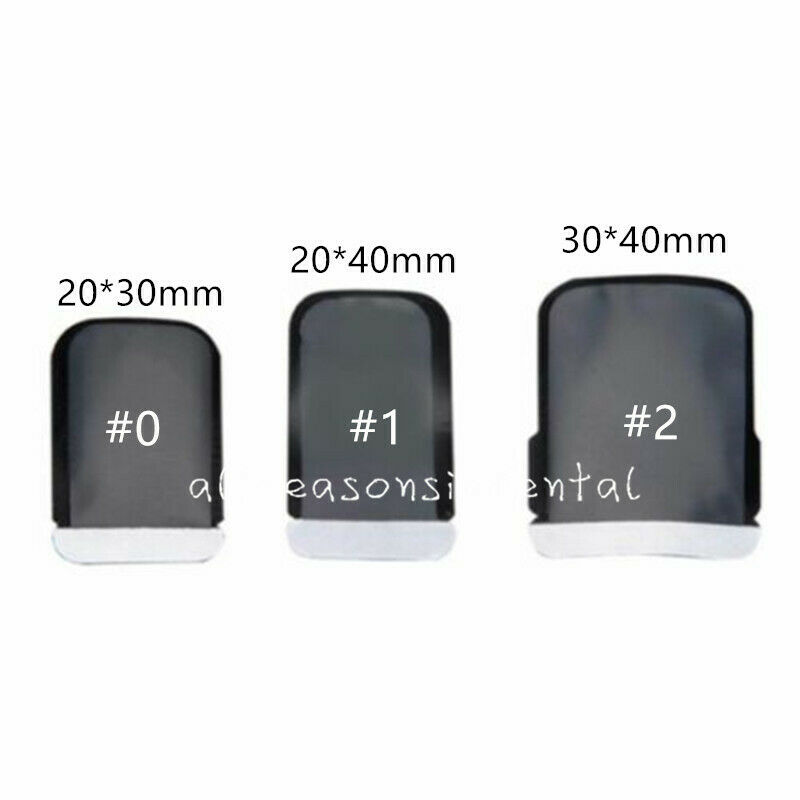
Acteon Sopro Life Dental Intraoral Camera, Cables, Manual, Docking Box and DVD
$ 1055.47
- Description
- Size Guide
Description
Lightly used but in perfect working condition. SN S900-7122-N.Excellent Images! Four different focus modes.
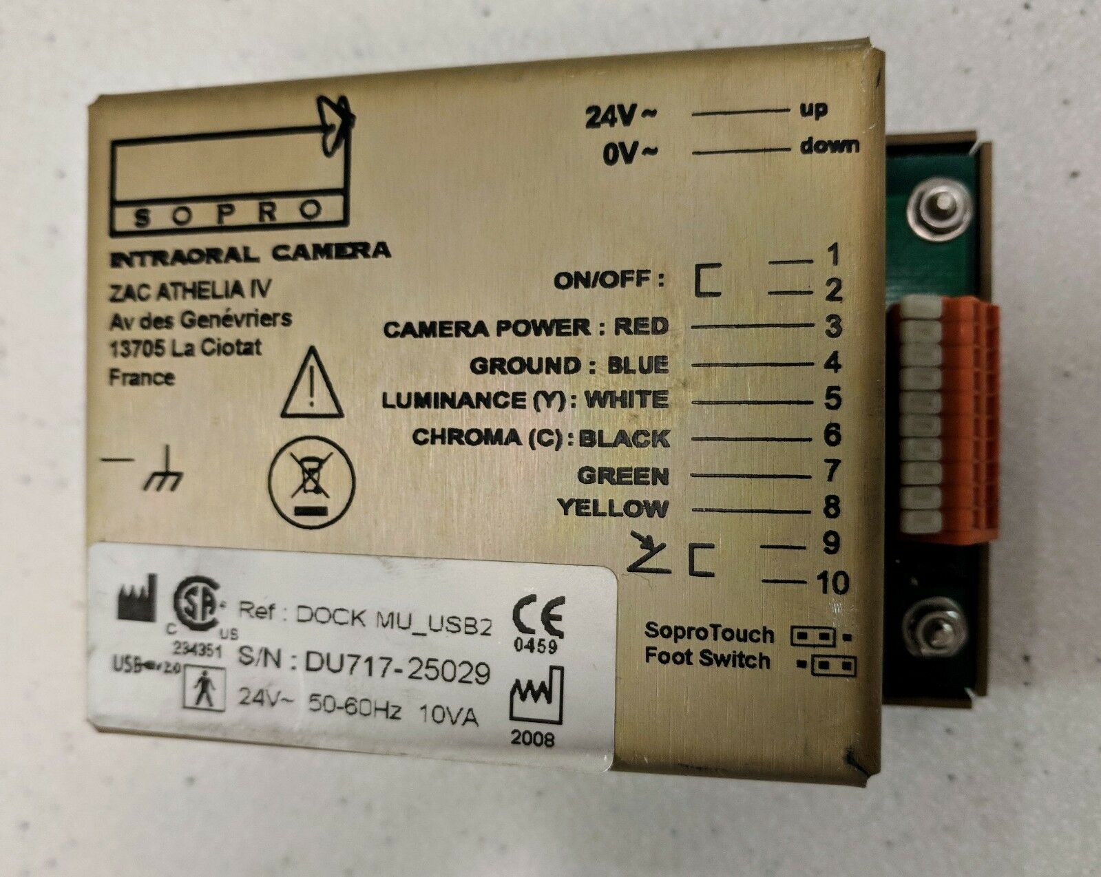
DOCK MU _USB2 S/N: DU717-25029
(We moved offices and eliminated one of our dental chairs.)
This camera was installed integrated into an a-dec 500 dental chair.
All sales are final, please ask any questions you have BEFORE purchasing.
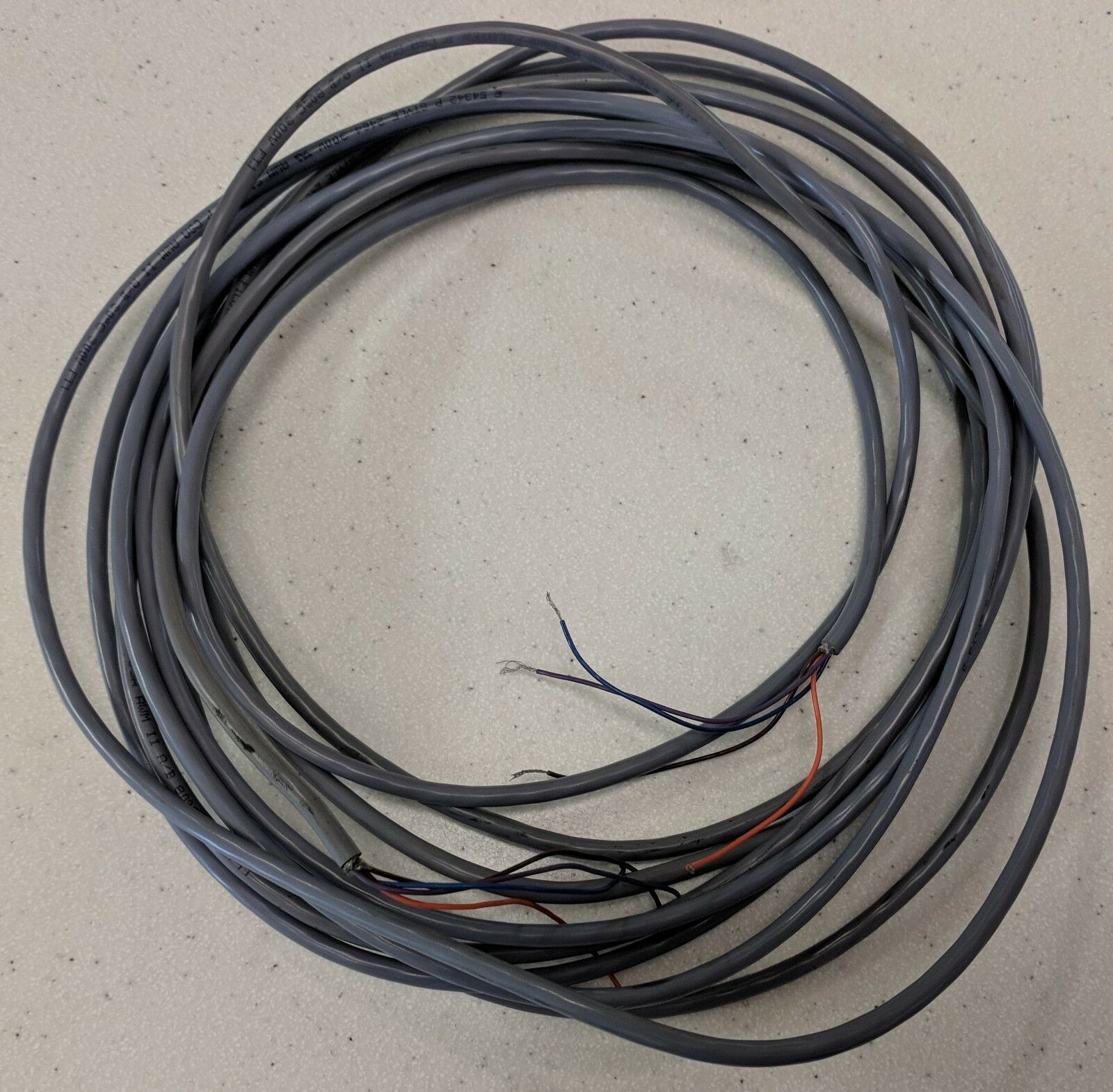
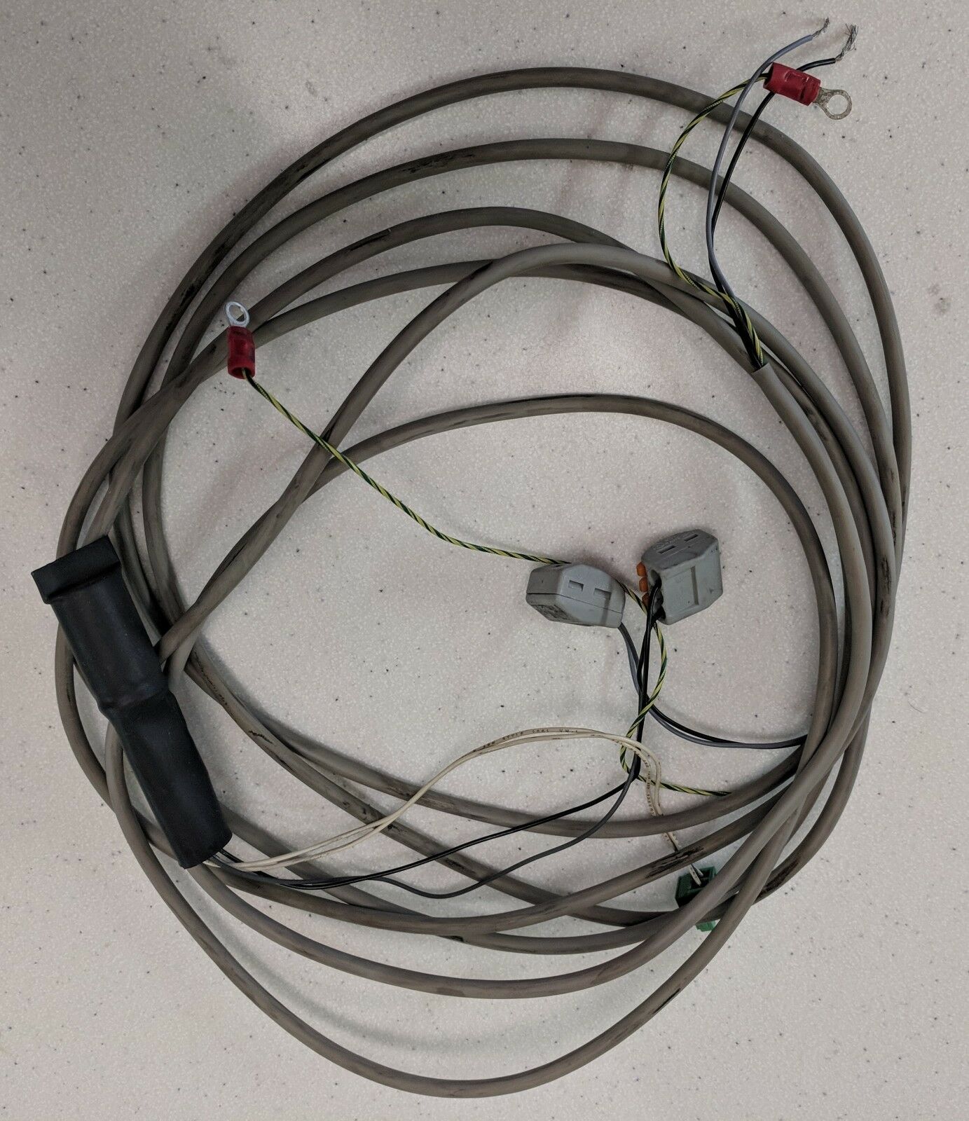
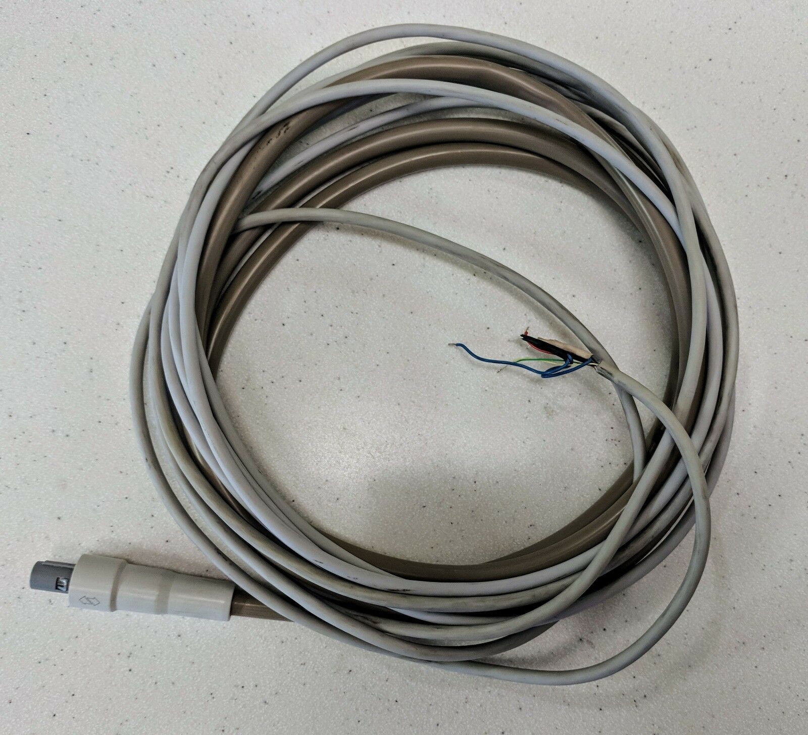
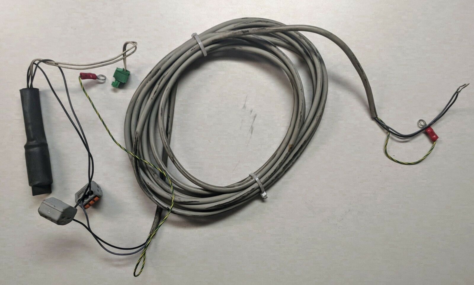
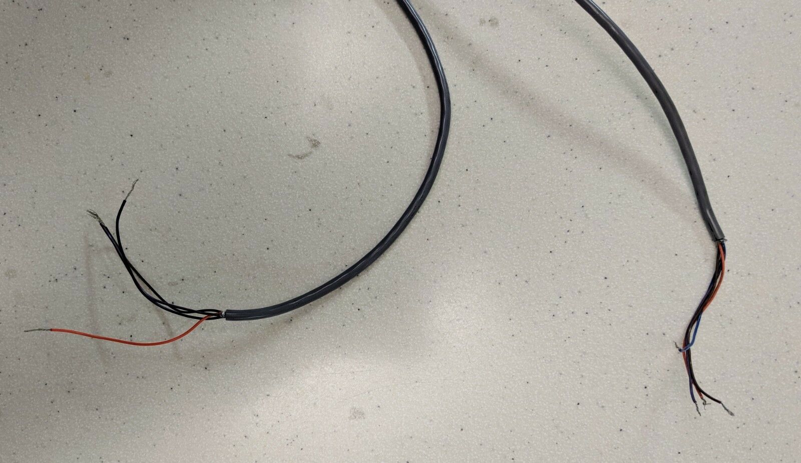
Includes all cables, manuals, disc and gold box pictured in listing photos.
SOPROLIFE
THE BLUE REVOLUTION
Dentistry will be changed forever… SOPROLIFE will allow you to see what was once invisible to your eyes.
SOPROLIFE offers you the ability to detect tooth decay at different stages of its development thus allowing you to determine the most adequate treatment.
The auto fluorescence technology in SOPROLIFE will allow you to detect occlusal or interproximal decay - even in its earliest stages.
SOPROLIFE allows you to differentiate healthy from infected tissue in order to excavate only the tissue which is diseased.
01
PRODUCT BENEFITS
DIAGNOSIS AID MODE
SOPROLIFE
®
is more accurate in identifying the development of occlusal and/or proximal carious lesions.
This mode potentially speeds up the decision making process in treatment planning and enables safer options for the patients by possibly reducing the number of x-rays.
TREATMENT AID MODE
Clinical performance is enhanced as SOPROLIFE enables you to visually differentiate between infected and affected tissue in the excavated site.
DAYLIGHT MODE
In white light, from Portrait to Macro, SOPROLIFE produces unequalled quality of image.
This mode not only enables you to communicate more effectively with your patients, but also gives you the ability to see details which are invisible to the naked eye.
02
MAIN CHARACTERISTICS
- A revolutionary concept which offers you two different views
- Save time by making a faster, accurate diagnosis
- Protect your patients by reducing the number of necessary X-rays
- Differentiate between healthy and infected tissue
- Enhance your clinical performance
07
CLINICAL CASE
INTERPROXIMAL CARIES OF STAGE 3 | MAN | 30 YEARS
INTERPROXIMAL CARIES OF STAGE 4 | MAN | 29 YEARS
PROXIMAL CARIES STAGE 2 | WOMAN | 35 YEARS
OCCLUSAL CARIES STAGE 2 | GIRL | 13 YEARS
Précédent
Suivant
Précédent
Suivant
The carious lesion is not visible under white light
The fluorescence image shows a demineralization on the marginal mesial ridge compared to the opposite ridge.
The radiography confirms the presence of the carious lesion of stage 3.
Under white light, the entry point of the caries and the cavity can be seen.
